6. Spatial attention in movement preparation
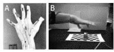
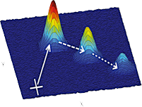
In psychophysical experiments we use secondary letter discrimination tasks to test how attention is deployed in the scene, e.g. while the participants prepare for actions. Often the primary tasks involve movement sequences such as series of saccades or pointing movements. We showed that if participants were asked to prepare for rapid movement sequences attention splits into multiple foci as to cover several (up to three) subsequent goal positions in parallel. Also if two movements are simultaneously prepared, e.g. when reaching bimanually, attention splits up between the intended points of application.
7. Encoding of sequences in the parietal and frontal cortex.
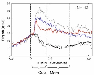
We recorded from single neurons in the parietal cortex while animals were preparing for a double-reach task to two peripheral goal locations. After mapping the response fields of individual cells in the parietal reach region (PRR) we placed either the first or the second goal of a reach sequence in the response field of the cell. This allowed us to attribute the cells‘s planning activity during a delay period to the representation of either the first or second movement goal. Most of the cells within PRR encoded immediate goals and subsequent goals equally well. This implies this part of parietal cortex encodes multiple movement goals of a planned hand movement sequence in parallel. Given that the parietal cortex has a roughly retinotopic organization, we argue that the eye-centered planning activity in PRR is a likely source of attentional top-down signals that facilitate visual processing at multiple goal positions. Functionally, the output of neural populations in PPC could be projected bi-directionally: the encoding of movement intention could be passed to further motor-related structures in frontal brain, whereas the very same output could also be back-projected (top-down) to early visual areas and subserve attentional selection-for-action.
8. Probing visual attention in human electrophysiology
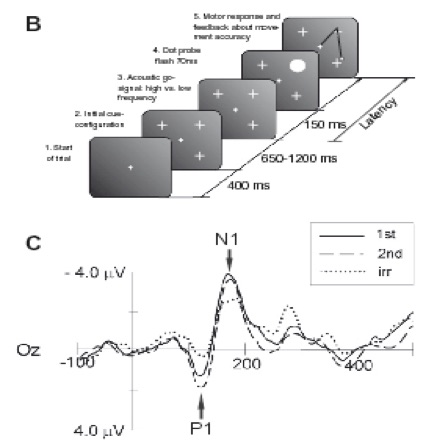
In order to probe the facilitation of visual input at future movement goals we asked human participants to prepare rapid sequences of reaches to peripheral goal locations. While keeping their eyes fixated at the centre of the screen, the subjects‘ visual attention was probed by analyzing brain potentials in the EEG that were evoked by task-irrelevant visual transients (so called ,dot-probes‘). These visual transients elicited larger P1/N1 components in the ERP signal if they happened to appear at the first or second intended goal location. Hence, also movement goals that lie in the rather remote future (i.e. at least some couple of hundreds milliseconds ahead) are visually pre-selected if a fluent and rapid motor response is required. The finding that multiple movement goals in a sequence are visually prepared in advance fits well with recent investigations in the chaining of movement components in everyday tasks like dish washing, tea making, etc. (see Hayhoe et al., 2003; Ballard et al., 1992).


1. Top-down mechanisms of spatial and non-spatial attention
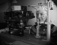
We use Magnetoencephalography (MEG) recordings to investigate top-down mechanisms of visual attention. The superb temporal resolution and the whole-head coverage of MEG allows us to study interactions of wide-spread neural networks by means of neural oscillations and synchrony. In particular, we became interested in mechanisms subserving non-spatial attention, e.g. when searching for a certain color.
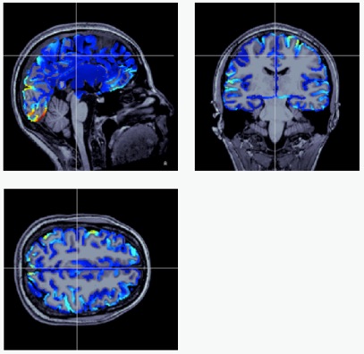
Within each subject we combine our temporally precise MEG recordings with spatially high-resoluting fMRI activity. In addition we confirm measures of functional connectivity (i.e., coherent neural oscillations in two or more areas) with the subject‘s individual anatomical connectivity by means of diffusion tractography (DTI).


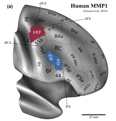
3. Frequency tagging functionally specialized brain areas
In MEG and EEG recording we often use frequency tagged stimuli: different aspects of a visual scene are updated periodically at slightly different presentation rhythms. After Fourier-transforming the whole brain activity, we can identify areas that picked up the stimulus‘ oscillation patterns and study their role in processing certain aspects of the layout. The movie on the right slides through Frequency domain (not time!) and shows activation peaks at two different stimulation frequencies evoked by overlaying dot patterns, one of which was attended.
4. Tagging the functional compartmentalization in high-level visual hierarchy
We developed a 2D-Fourier based frequency-tagging approach that allows tagging different aspects of a scene simultaneously and retrieving phase-locking values and information about the relative phase-lags (,latencies‘) for various functional compartmentalizations in high-level visual cortex.
5. Functional and anatomical connectivity
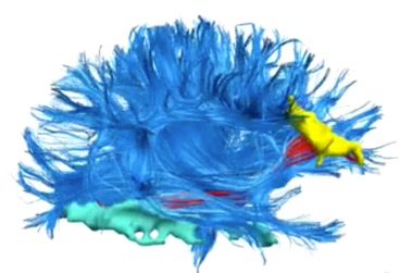
When we study patterns of functional connectivity, e.g. in terms of coherent oscillatory activity in two brain regions, we also try to to pinpoint the anatomical basis of these functional connectivities. To do so we analyze DTI scans in our participants and reconstruct fiber bundles in the white matter that connect the two sites. In the movie to the right you see in red the fiber bundles that connect two functionally coupled brain regions: the inferior-frontal cortex (yellow) and IT cortex (green).
Daniel Baldauf
CIMeC - Center for Mind and Brain Sciences
University of Trento

Human Imaging Center
Via delle Regole, 101
38100 Mattarello (TN), Italy

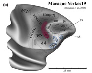
2. Structure, function and connectivity fingerprints of prefrontal cortex:
The human prefrontal cortex contains two prominent areas, the frontal eye field and the inferior frontal junction, that are crucially involved in the orchestrating functions of attention, working memory and cognitive control. Motivated by comparative evidence in non-human primates, we review the
human neuroimaging literature, suggesting that the functions of these regions can be clearly dissociated. We found remarkable differences in how these regions relate to sensory domains and visual topography, top-down and bottom-up spatial attention, spatial versus non-spatial (i.e., feature- and object-based) attention and working memory and, finally, the multiple demand system. Functional magnetic resonance imaging (fMRI) studies using multivariate pattern analysis reveal the selectivity of the frontal eye field and inferior frontal junction to spatial and non-spatial information, respectively. The analysis of functional and effective connectivity provides evidence of the modulation of the activity in downstream visual areas from the frontal eye field and inferior frontal junction and sheds light on their reciprocal influences.
We work on further characterizing their involvement in a spatial (‘ where’ ) and a non-spatial (‘ what’ ) network, respectively, highlighting segregated brain networks that allow biasing visual selection and working memory performance to support goal-driven
behavior.